Super resolution scanning laser microscopy and ophthalmoscopy
High-resolution microscopy is crucial for accurate diagnosis of medical diseases and has revolutionized the field of biological sciences. However, conventional microscopes have a light diffraction limit that limits their spatial resolution. Structured illumination has been explored to overcome this limit in wide field light microscopy, but practical deployment of structured illumination microscopy (SIM) for in vivo retinal imaging is often challenging due to phase errors caused by eye movement. To address this, our team developed super-resolution scanning laser microscopy using virtually structured detection (VSD), which provides an easy, low-cost, and phase-artifact free strategy to achieve super-resolution in scanning laser microscopy. We have demonstrated the feasibility of in vivo super-resolution scanning laser ophthalmoscopy (SLO) by combining a line-scanning strategy. Our ongoing research investigates the potential of using VSD-based SLO for in vivo super-resolution functional imaging of human photoreceptors, with significant implications for improving the diagnosis and treatment of retinal diseases.
Figure Heading link
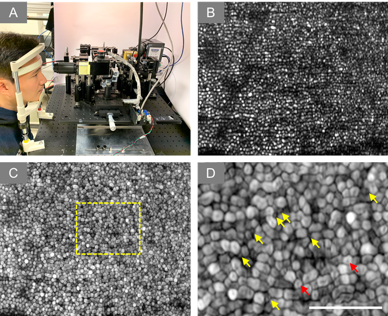
Figure. (A) Experiment setup of super-resolution SLO. (B) Representative image of human retina. (C) Dynamic motility analysis of retinal photoreceptors. (D) Enlarged illustration of the yellow window in C. The yellow and red arrowheads point to individual rod and cone photoreceptors, respectively.
Current development and future direction Heading link
Animal study
Our team has been working on virtually structured detection (VSD) technology to improve the resolution of conventional scanning laser microscopy (SLM), which has resulted in the resolution of single photoreceptor cells. By combining VSD with line-scanning microscopy (LSM), we achieved increased imaging speed and resolution, despite the challenges of modulating illumination and detection light for ophthalmic applications. By using high-speed cameras, we were able to create VSD-based super-resolution images that reveal individual photoreceptors and nerve fiber bundles with improved image contrast and signal-to-noise ratio. Our success in this area led us to develop a virtually structured detection-based super-resolution ophthalmoscope, which achieved subcellular spatial resolution and millisecond temporal resolution for in vivo imaging of transient receptor potential (TRP). This allowed us to study the spatiotemporal properties of TRP under variable light intensity stimuli and provided evidence of a correlation between TRP and the early stages of phototransduction.
Human study
In 2021, our team achieved a significant breakthrough in VSD technology by developing a technique that improves resolution and enables the clear visualization of individual retinal photoreceptors without the need for expensive and complex adaptive optics (AO) equipment. This is particularly significant because AO is often not practical in certain clinical settings. Additionally, we introduced a dynamic motility processing technique that enhances the image contrast of individual photoreceptors, making it easier to distinguish between rod and cone photoreceptors. These advances have the potential to greatly improve our understanding of retinal photoreceptor function and pathology, leading to improved diagnostic and treatment options for retinal diseases.
Future directions
Our team aims to optimize the VSD technology with customized optical design to improve the imaging performance, which can lead to a cost-effective method for clinical deployments of high-resolution ophthalmoscopy. We are also exploring the feasibility of combining VSD and adaptive optics (AO) to break the diffraction limit and achieve diffraction-limited resolution, which could further advance retinal study and eye disease management.


Related publications Heading link
2021
- Yao, Xincheng, Rongwen Lu, Benquan Wang, Yiming Lu, and Tae-Hoon Kim. Super-resolution ophthalmoscopy: Virtually structured detection for resolution improvement in retinal imaging. Exp Biol Med (Maywood) 2021;246:249-59 [Full text]
- Lu, Yiming, Taeyoon Son, Tae-Hoon Kim, David Le, and Xincheng Yao, “Virtually structured detection enables super-resolution ophthalmoscopy of rod and cone photoreceptors in human retina,” Quant Imaging Med Surg 2021 [Full text]
2018
- Lu, Yiming, Changgeng Liu, and Xincheng Yao. “In vivo super-resolution imaging of transient retinal phototropism evoked by oblique light stimulation.” Journal of biomedical optics 23, no. 5 (2018): 050502-050502.[Full Text]
2017
- Liu, Changgeng, Damber Thapa, and Xincheng Yao. “Digital adaptive optics confocal microscopy based on iterative retrieval of optical aberration from a guidestar hologram.” Optics Express 25, no. 7 (2017): 8223-8236. [Full Text]
2016
- Liu, Changgeng , Yanan Zhi, Benquan Wang, Damber Thapa, Yanjun Chen, Minhaj Alam, Yiming Lu, and Xincheng Yao. “In vivo super-resolution retinal imaging through virtually structured detection.” Journal of biomedical optics 21, no. 12 (2016): 120502-120502.[Full Text]
2015
- Zhi, Yanan, Benquan Wang, and Xincheng Yao. “Super-resolution scanning laser microscopy based on virtually structured detection.” Critical Reviews™ in Biomedical Engineering 43, no. 4 (2015). [Full Text]
- Zhi, Yanan, Rongwen Lu, Benquan Wang, Qiuxiang Zhang, and Xincheng Yao. “Rapid super-resolution line-scanning microscopy through virtually structured detection.” Optics letters 40, no. 8 (2015): 1683-1686.[Full Text]
2013
- R.W. Lu, B.Q. Wang, Q.X. Zhang, and X.C. Yao. Super-resolution scanning laser microscopy through virtually structured detection, Biomedical Optics Express 4, 1673-1682 (2013) [Full Text]