Wide field fundus photography
Wide field fundus photography is essential for remote screening, diagnosis, and treatment evaluation of eye diseases. However, the complexity of conventional transpupillary illumination and imaging mechanisms has made it technically challenging to construct a wide field fundus camera. To overcome this challenge, we have developed ultra-widefield fundus cameras that use trans-palpebral and trans-pars-planar illumination techniques. Additionally, we have developed a novel miniaturized indirect ophthalmoscopy approach for wide field fundus photography. By further developing low-cost and easy-to-use widefield fundus photography, we can offer an affordable solution for telemedicine assessment of eye diseases, thereby improving access to eye care for patients in rural and underserved areas. Our team of experienced researchers is dedicated to using the latest technology and cutting-edge techniques to develop and refine wide field fundus photography for both adult and pediatric patients.
Figure Heading link
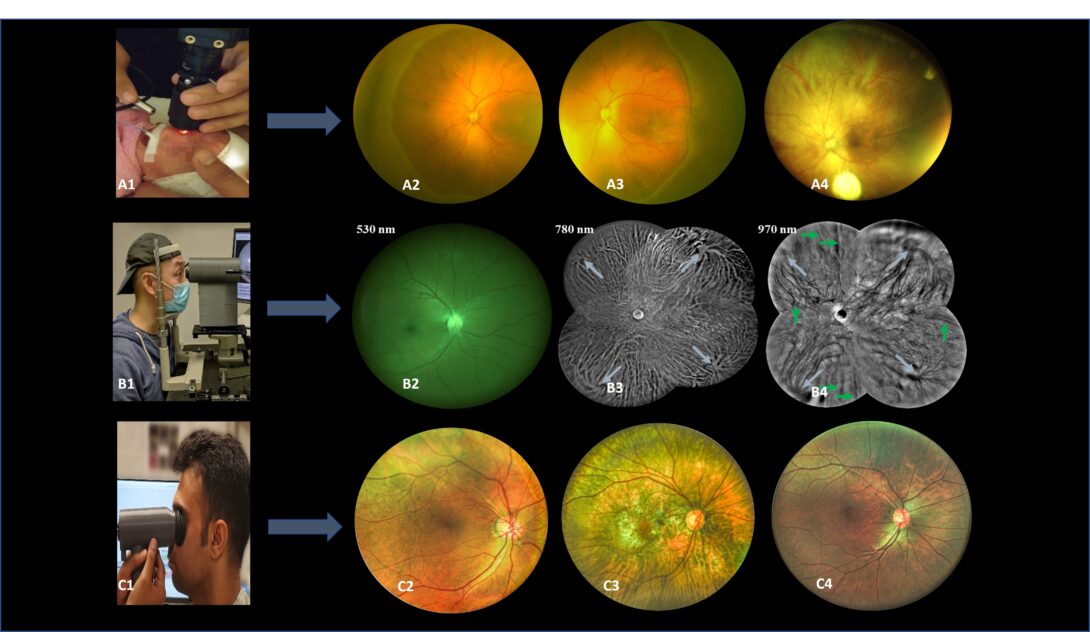
Figure1 A1. Pediatric fundus camera during imaging. A2-A4. Sample images from pediatric fundus camera. B1. Trans eyelid fundus camera during imaging. B2-B4. Sample images with different light wavelengths from the trans-eyelid fundus system. C1. Miniaturized HDR trans pupillary fundus camera during imaging. C2-C4. Sample HDR images from trans pupillary fundus system.
Current development and future direction Heading link
Ultra-widefield fundus photography
Our research team has been focused on the development of ultra-widefield fundus cameras that utilize the trans-pars-plana illumination technique to achieve a 200° field of view for both adults and pediatrics without pupil dilation. This advancement enables easy examination of the retina and improves the ability to detect and diagnose a variety of eye diseases. Additionally, we have also developed a non-contact 140° wide field fundus camera that utilizes trans-palpebral illumination. In addition to expanding the field of view, we have also been exploring the use of multispectral fundus imaging to visualize both the retinal and choroidal fundus images. We have been experimenting with color balancing techniques to enhance the image quality of fundus images. Furthermore, we have also made efforts to create portable fundus imaging modalities by using benchtop systems and smartphone-based devices. These innovations have the potential to improve access to eye care and enable remote screening and diagnosis of eye diseases.
Miniaturized indirect ophthalmoscopy
Our laboratory has developed a series of innovative imaging techniques to advance the field of ophthalmology, including miniaturized indirect ophthalmoscopy for wide field fundus photography. Our first prototype was a low-cost, portable, wide field smartphone fundus camera that utilized miniaturized indirect ophthalmoscopy illumination. We then improved upon this design by using a rotation stage to remove reflection artifacts and expand the field of view. We also incorporated high dynamic range (HDR) imaging capability for better image quality. To eliminate illumination reflectance artifacts, we utilized orthogonal polarization control. These advancements have resulted in a portable, nonmydriatic, wide field fundus camera that can be used for both adults and pediatric patients.
Future directions
Our laboratory is focused on continuously improving the optical design of the fundus cameras to expand the field of view and enhance image quality. In addition, we are developing multimodal fundus imaging modalities that provide various types of retinal information to improve the detection and prolonged follow-up of eye diseases. Our approach includes not only obtaining structural information but also incorporating functional information to enhance disease diagnosis. By combining multiple imaging techniques, we can extract a more comprehensive set of biomarkers for a variety of eye diseases, providing clinicians with an unprecedented level of insight into their patients’ eye health.


Related publications Heading link
2024
- Mojtaba Rahimi, Alfa Rossi, Taeyoon Son, Albert K. Dadzie, Behrouz Ebrahimi, Mansour Abtahi, Michael J. Heiferman, Xincheng Yao; Multispectral Fundus Photography of Choroidal Nevi With Trans-Palpebral Illumination. Trans. Vis. Sci. Tech. 2024;13(3):25.[Full text]
- Rossi, Alfa, Yushun Zeng, Mojtaba Rahimi, Taeyoon Son, Michael J. Heiferman, Chen Gong, Xin Sun, Mohammad Soleimani, Ali R. Djalilian, Mark S. Humayun, and et al. 2024. “Integrating a Fundus Camera with High-Frequency Ultrasound for Precise Ocular Lesion Assessment” Biosensors 14, no. 3: 127. https://doi.org/10.3390/bios14030127.[Full text]
2023
- Rossi, Alfa, Mojtaba Rahimi, Taeyoon Son, R. V. Paul Chan, Michael J. Heiferman, and Xincheng Yao, “Preserving polarization maintaining photons for enhanced contrast imaging of the retina,” Biomed. Opt. Express 14, 5932-5945 (2023).[Full text]
- Rahimi, Mojtaba, Alfa Rossi, Taeyoon Son, Devrim Toslak, David Le, Mansour Abtahi, Michael J. Heiferman, R. V. Paul Chan, and Xincheng Yao, “Evaluating spatial dependency of the spectral efficiency in trans-palpebral illumination for widefield fundus photography,” Biomed. Opt. Express 14, 5629-5641 (2023).[Full text]
- Rossi, Alfa, Mojtaba Rahimi, David Le, Taeyoon Son, Michael J. Heiferman, RV Paul Chan, and Xincheng Yao. “Portable widefield fundus camera with high dynamic range imaging capability.” Biomedical Optics Express 14, no. 2 (2023): 906-917. [Full text]
2022
- Son, Taeyoon, Jiechao Ma, Devrim Toslak, Alfa Rossi, Hoonsup Kim, R. V. Chan, and Xincheng Yao. “Light color efficiency-balanced trans-palpebral illumination for widefield fundus photography of the retina and choroid.” Scientific Reports 12, no. 1 (2022): 1-11. [Full text]
- Yao, Xincheng, Taeyoon Son, and Jiechao Ma. “Developing portable widefield fundus camera for teleophthalmology: Technical challenges and potential solutions.” Experimental Biology and Medicine (2021): 15353702211063477. [Full text]
2021
- Yao, Xincheng, Devrim Toslak, Taeyoon Son, and Jiechao Ma, “Understanding the relationship between visual-angle and eye-angle for reliable determination of the field-of-view in ultra-wide field fundus photography,” Biomed. Opt. Express 12, 6651-6659 (2021). [Full text]
2020
- Toslak, Devrim, Taeyoon Son, Muhammet Kazim Erol, Hoonsup Kim, Tae-Hoon Kim, R. V. Paul Chan, and Xincheng Yao, “Portable ultra-widefield fundus camera for multispectral imaging of the retina and choroid,” Biomed. Opt. Express 11, 6281-6292 (2020) [Full text].
- Toslak, Devrim, Felix Chau, Muhammet Kazim Erol, Changgeng Liu, R. V. Paul Chan, Taeyoon Son, and Xincheng Yao, “Trans-pars-planar illumination enables a 200° ultra-wide field pediatric fundus camera for easy examination of the retina,” Biomed. Opt. Express 11, 68-76 (2020) [Full text].
2018
- Wang, Benquan, Devrim Toslak, Minhaj Nur Alam, R. V. Chan, and Xincheng Yao. “Contact-free trans-pars-planar illumination enables snapshot fundus camera for nonmydriatic wide field photography.” Scientific Reports 8, no. 1 (2018): 1-9.[Full Text]
- Toslak, Devrim, Changgeng Liu, Minhaj Nur Alam, and Xincheng Yao. “Near-infrared light-guided miniaturized indirect ophthalmoscopy for nonmydriatic wide field fundus photography.” Optics letters 43, no. 11 (2018): 2551-2554. [Full Text]
- Toslak, Devrim, Ali Ayata, Changgeng Liu, Muhammet Kazim Erol, and Xincheng Yao. “Wide field smartphone fundus video camera based on miniaturized indirect ophthalmoscopy.” Retina (Philadelphia, Pa.) 38, no. 2 (2018): 438. [Full Text]
2017
- Toslak, Devrim, Damber Thapa, Muhammet Kazim Erol, Yanjun Chen, and Xincheng Yao. “Smartphone-based imaging of the corneal endothelium at sub-cellular resolution.” Journal of modern optics 64, no. 12 (2017): 1229-1232. [Full Text]
2016
- Toslak, Devrim, Damber Thapa, Yanjun Chen, Muhammet Kazim Erol, RV Paul Chan, and Xincheng Yao. “Trans-palpebral illumination: an approach for wide-angle fundus photography without the need for pupil dilation.” Optics letters 41, no. 12 (2016): 2688-2691. [Full Text]